Brachial Plexus
Description & Overview
1. Before You Start
Before studying the brachial plexus, revise these basic concepts:
- Spinal nerves: Nerves formed by the union of the anterior (ventral) root and posterior (dorsal) root of the spinal cord.
[Read more] | [Watch video] - Anterior rami (ventral rami): Branches of spinal nerves that supply the anterolateral trunk and, through nerve plexuses, most of the limbs. The brachial plexus is formed by the anterior rami of C5–T1 spinal nerves.
[Read more] | [Watch video] - Motor supply (efferent supply): Nerve fibres that carry signals from the central nervous system to muscles, producing movement.
[Read more] | [Watch video] - Sensory supply (afferent supply): Nerve fibres that carry information such as touch, pain, and temperature from the body to the central nervous system.
[Read more] | [Watch video] - Peripheral nerves: Nerves outside the brain and spinal cord that carry motor, sensory, and sometimes autonomic fibres.
[Read more] | [Watch video] - Upper limb anatomy: The brachial plexus supplies most of the muscles and skin of the upper limb, including the shoulder, arm, forearm, and hand.
[Read more] | [Watch video]
2. Topic Overview
The brachial plexus is a network of nerves formed mainly by the anterior rami of the lower four cervical spinal nerves and the first thoracic spinal nerve, C5–T1. It provides motor and sensory innervation to most of the upper limb.
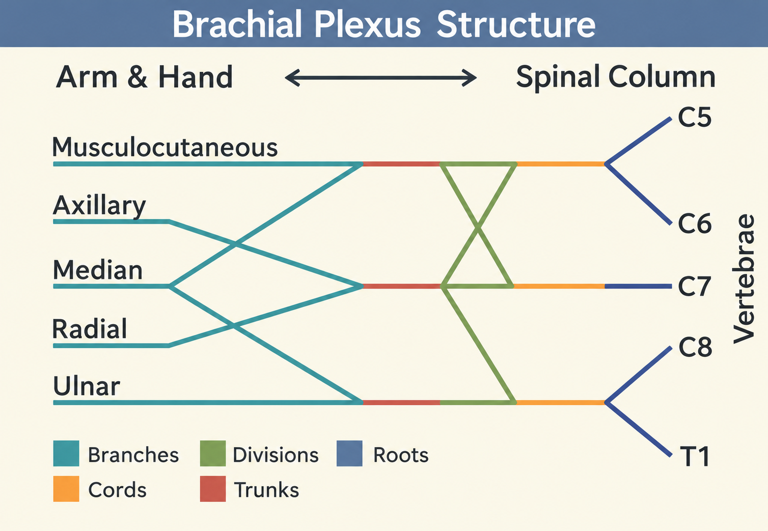
The plexus begins in the lower part of the neck, passes between the anterior and middle scalene muscles, enters the axilla, and gives rise to the major nerves of the upper limb.
Its main arrangement is:
Roots → Trunks → Divisions → Cords → Branches
The five roots form three trunks. Each trunk divides into an anterior and a posterior division. These six divisions reorganize into three cords, which give rise to the terminal branches and several collateral branches.
The five major terminal nerves are the musculocutaneous, axillary, radial, median and ulnar nerves. These nerves supply muscles and skin in different regions of the upper limb.
3. Understand the Topic
Want a simpler Hindi-English explanation? Read this topic in Hinglish.
A. Formation of the Brachial Plexus
The brachial plexus is formed mainly by the anterior rami of spinal nerves C5, C6, C7, C8 and T1.Cervical Nerve Anatomy Animation
Remember that C5–T1 refers to spinal nerve roots contributing to the plexus, not to the five terminal nerves.
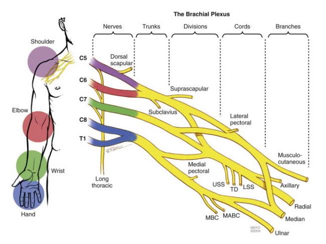
1. Roots
The five roots are:
- C5
- C6
- C7
- C8
- T1
They are the anterior rami of the corresponding spinal nerves.
Important branches from the roots:
- Dorsal scapular nerve (mainly C5): Supplies the rhomboid muscles and usually levator scapulae.
- Long thoracic nerve (C5–C7): Supplies serratus anterior.
2. Trunks
The roots combine to form three trunks:
| Trunk | Formation |
|---|---|
| Upper trunk | C5 + C6 |
| Middle trunk | Continuation of C7 |
| Lower trunk | C8 + T1 |
Branches from the upper trunk:
- Suprascapular nerve (C5–C6): Supplies supraspinatus and infraspinatus.
- Nerve to subclavius (C5–C6): Supplies subclavius.
3. Divisions
Each trunk divides into an anterior and a posterior division, giving six divisions in total.
- Anterior divisions: Mainly contribute to nerves supplying the anterior compartments of the upper limb, including many flexor muscles.
- Posterior divisions: Mainly contribute to nerves supplying the posterior compartments, including many extensor muscles.
This is a useful general pattern, but it is not an absolute rule for every muscle or nerve.
The divisions do not usually give off major named branches before they form the cords.
4. Cords
The six divisions combine to form three cords. Their names are based on their position relative to the second part of the axillary artery.
| Cord | Formation |
|---|---|
| Lateral cord | Anterior divisions of upper and middle trunks |
| Medial cord | Anterior division of lower trunk |
| Posterior cord | All three posterior divisions |
Branches from the lateral cord:
- Lateral pectoral nerve (C5–C7)
- Musculocutaneous nerve (C5–C7)
- Lateral root of median nerve (C5–C7)
Branches from the medial cord:
- Medial pectoral nerve (C8–T1)
- Medial cutaneous nerve of arm (C8–T1)
- Medial cutaneous nerve of forearm (C8–T1)
- Ulnar nerve (mainly C8–T1)
- Medial root of median nerve (C8–T1)
Branches from the posterior cord:
- Upper subscapular nerve (C5–C6)
- Thoracodorsal nerve (C6–C8)
- Lower subscapular nerve (C5–C6)
- Axillary nerve (C5–C6)
- Radial nerve (C5–T1)
5. Terminal Branches
The five major terminal nerves are:
| Nerve | Root values | Main motor supply | Important sensory area | |
|---|---|---|---|---|
| Musculocutaneous | C5–C7 | Anterior compartment of arm: coracobrachialis, biceps brachii and brachialis | Lateral side of forearm through the lateral cutaneous nerve of forearm | |
| Axillary | C5–C6 | Deltoid and teres minor | Skin over the upper lateral arm, including the regimental badge area | |
| Radial | C5–T1 | Extensor muscles of arm and forearm; also supplies triceps | Mainly posterior arm and forearm and part of the dorsolateral hand | |
| Median | C5–T1 | Most forearm flexors and pronators; thenar muscles and lateral two lumbricals | Lateral palm and palmar surface of the lateral 3½ digits, with distal dorsal parts of these digits | |
| Ulnar | Mainly C8–T1 | Most intrinsic hand muscles; flexor carpi ulnaris and medial half of flexor digitorum profundus | Medial 1½ digits and adjacent medial hand on palmar and dorsal surfaces |
Important clarification: Root values can vary slightly. Root values may vary slightly between individuals because the pattern of nerve connections can differ from person to person. The values given represent the usual pattern.
Remember: Learn the usual root values first because they represent the standard anatomical pattern. The variations are important for understanding...
✨ Reading in Preview Mode (50%)
Join PhysioSource to unlock full detailed descriptions, diagrams, and clinical insights.
Login to ContinueLearning Materials
Model Question Bank - Brachial Plexus
MNEMONICS FOR THE BRACHIAL PLEXUS
ORGANIZATION
Mnemonic: Read The Diagram Carefully, Bro
* R — Roots
* T — Trunks
* D — Divisions
* C — Cords
* B — Branches
LATERAL CORD BRANCHES
Mnemonic: Low-Level Math
* L — Lateral pectoral nerve
* L — Lateral root of median nerve
* M — Musculocutaneous nerve
MEDIAL CORD BRANCHES
Mnemonic: M4U (4 M's and 1 U)
* M — Medial cutaneous nerve of arm
* M — Medial cutaneous nerve of forearm
* M — Medial pectoral nerve
* M — Medial root of median nerve
* U — Ulnar nerve
POSTERIOR CORD BRANCHES
Mnemonic: ULTRA
* U — Upper subscapular nerve
* L — Lower subscapular nerve
* T — Thoracodorsal nerve
* R — Radial nerve
* A — Axillary nerve
NERVE LESIONS
Mnemonic: DR. CUMA (Deformity paired with the injured nerve)
* DR — Drop wrist \ Radial nerve lesion
* CU — Claw hand \ Ulnar nerve lesion
* MA — Median nerve lesion \ Ape hand
Bottom Line
KEY TAKEAWAY The brachial plexus is a network of nerves formed mainly by the anterior rami of C5–T1. It supplies most of the muscles and skin of the upper limb. Its arrangement into roots, trunks, divisions, cords and branches helps explain the distribution of its nerves and the effects of nerve injuries. QUICK REVISION POINTS - ROOTS: C5, C6, C7, C8 and T1. - TRUNKS: Upper (C5–C6), middle (C7) and lower (C8–T1). - DIVISIONS: Each trunk divides into anterior and posterior divisions. - CORDS: Lateral, medial and posterior; named in relation to the second part of the axillary artery. - FIVE MAJOR TERMINAL NERVES: Musculocutaneous, axillary, radial, median and ulnar. - ROOT BRANCHES: Dorsal scapular and long thoracic nerves. - UPPER TRUNK BRANCHES: Suprascapular nerve and nerve to subclavius. - POSTERIOR CORD BRANCHES: Upper subscapular, thoracodorsal, lower subscapular, axillary and radial nerves. - UPPER TRUNK INJURY: Commonly associated with Erb-Duchenne palsy. - LOWER TRUNK INJURY: Commonly associated with Klumpke palsy. - LONG THORACIC NERVE INJURY: Can cause winging of the scapula. EXAM PERSPECTIVE For a long-answer question, begin with the definition and root values. Explain the formation of the roots, trunks, divisions and cords, then describe the branches at each level. Include the five terminal nerves, their root values, motor and sensory distributions, and clinical importance. Practise drawing a labelled brachial plexus diagram. Make sure the roots, trunks, divisions, cords and terminal branches are clearly shown. For short-answer questions, prepare the branches of each cord and the clinical features of upper and lower trunk injuries. CLINICAL INSIGHT The level of a brachial plexus injury influences which muscles become weak and which skin regions lose sensation. An upper trunk injury may affect shoulder and elbow movements, while a lower trunk injury may particularly affect hand function. Injury to individual terminal nerves produces more localized patterns, such as wrist drop in some radial nerve injuries or scapular winging after long thoracic nerve injury.